About
How interventional radiology is changing what is possible for patients
- Home
- About
- Latest news
- How interventional radiology is changing what is possible for patients
31 August 2026
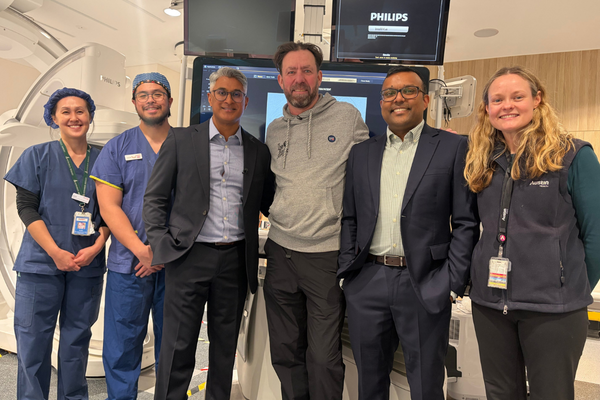
When Luke Johnston arrived at Austin Hospital struggling to breathe, scans revealed large blood clots in his lungs.
His condition was serious. For someone in Luke’s situation, with what is known as an intermediate to high-risk pulmonary embolism, the risk of dying can be around one in 10.
But rather than major surgery, our Interventional Radiology (IR) team was able to remove the clots through a tiny incision while Luke remained awake.
It is one example of just how far Interventional Radiology has come.
Using medical imaging to guide them, interventional radiologists perform minimally invasive procedures through small incisions, navigating through blood vessels and other parts of the body to reach exactly where treatment is needed.
At Austin Health, the IR team provides a 24-hour service across emergency and planned care, working closely with teams including Respiratory Medicine, the Emergency Department, Intensive Care and Surgery.
Dr Dinesh Ranatunga, Head of Interventional Radiology, said advances in the field are changing what is possible for patients.
“We have seen iterations of this technology over the last couple of decades, but it is really in the last five years that we have seen the technology improve in leaps and bounds,” Dinesh said.
“We can now do these procedures much more rapidly and safely, and help patients get home much sooner.”
One of those advances is the ability to remove large blood clots directly from the lungs of some patients with a pulmonary embolism. Austin Health began performing these procedures in mid-2024 and the team has now completed around 40 cases.
Interventional Radiologist Dr Sonal Udayasiri said the procedure provides another treatment option for patients with serious pulmonary embolisms who may otherwise face significant risks.
“The main strategy that we have for this is to watch and wait, which is to keep someone in close observation somewhere like intensive care and hope that with blood-thinning medication they improve,” Sonal said.
“If they do not improve, most of the treatments that we have, although possible, are very risky. To be able to offer something like this, which could be applied to a lot more people at a lower risk, is fantastic.”
For Luke, the first sign something was seriously wrong was increasing breathlessness.
He had spent several days in bed with influenza when his GP checked his oxygen levels and found they had fallen into the mid-80s. He was told to go straight to hospital.
By then, climbing a flight of stairs was enough to leave him feeling like he was going to pass out. Scans at Austin Hospital revealed the blood clots in his lungs.
“For the situation Luke was in, what we call an intermediate to high-risk pulmonary embolism, we would estimate about a one in 10 chance of dying from that condition,” Sonal said.
Sonal and the IR team were able to remove the clots using a minimally invasive procedure.
With Luke awake during his procedure, a tube was inserted through a small incision in his groin and guided through his blood vessels to the arteries in his lungs, allowing the team to remove the clots obstructing blood flow.
Luke spent the procedure listening to podcasts through his headphones.
“I did not want to hear what they were saying. I was just lying there listening to podcasts,” Luke said.
Two days later, he walked out of hospital.
“I was told to take six or eight weeks off work, and I was working in four days,” Luke said.
For Dinesh, cases like Luke’s demonstrate how much minimally invasive treatment has changed the options available to patients.
“Before minimally invasive techniques, this would either have to be treated with medical therapy, just thinning the blood, or sometimes clot-busting medications, which can have a high risk of bleeding,” he said.
“The alternative otherwise is to cut someone’s chest open. You can imagine having a massive cut and a big operation. We have moved well away from that.”
Sonal said the speed at which some patients can recover is one of the biggest benefits.
“A high-risk clot in the lungs could mean weeks of being in hospital, potentially even in intensive care on life support. With this procedure, we can potentially reduce that to patients walking out of hospital under their own power in days, sometimes even hours," he said.
While technology continues to advance, Sonal said the work relies on people and expertise across Austin Health.
For patients with serious pulmonary embolisms, specialists from Interventional Radiology, Respiratory Medicine, the Emergency Department and Intensive Care come together rapidly to decide the best treatment for each individual patient.
“The procedure itself is obviously a very cool thing, but it is the culmination of a lot of work across different departments and teams. That teamwork is probably the most critical part of what we do,” Sonal said.
Pulmonary embolisms are just one part of the IR team’s work.
The team uses minimally invasive techniques across emergency and planned care, including stopping serious bleeding, delivering cancer treatments and treating conditions including fibroids and enlarged prostates.
And Dinesh believes the field will continue to expand.
“As the evidence improves and we collect data on all these patients, the sky really is the limit with what we can do with minimally invasive techniques,” he said.
For Luke, learning just how serious his condition had been brought home the significance of the care he received.
“That makes you feel very, very fortunate. I’m just so grateful to the team.”